What is an Electron Microscope?
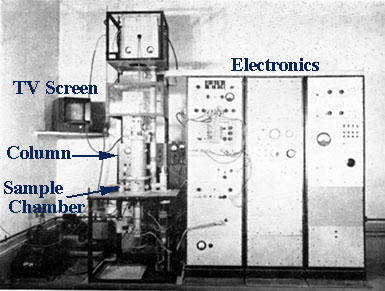
The development of the Scanning Electron Microscope in the early 1950’s brought with it new areas of study in the medical and physical sciences because it allowed examination of a great variety of specimens.
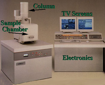
As in any microscope the main objective is for magnification and focus for clarity. An optical microscope uses lenses to bend the light waves and the lenses are adjusted for focus. In the SEM, electromagnets are used to bend an electron beam which is used to produce the image on a screen. By using electromagnets an observer can have more control in how much magnification he/she obtains. The electron beam also provides greater clarity in the image produced.

The SEM is designed for direct studying of the surfaces of solid objects.
